New study reveals role of cell surface sugars in neuronal navigation
24. 07. 2024
An international team of scientists from the Faculty of Science at Charles University, the Institute of Molecular Genetics of the Czech Academy of Sciences, and Stanford University has uncovered the molecular mechanisms by which cell surface sugars influence the navigation of neurons in the nervous system. Their results were published in the prestigious journal PNAS.
The adult human brain contains approximately 100 billion nerve cells, or neurons. Each neuron forms an average of 1,000 connections with other neurons, resulting in approximately 100 trillion synaptic contacts. These connections are crucial for our cognitive abilities, sensory perception, and memory. Understanding how such a vast network of contacts develops in the brain is a central question in neurobiology.
During the development of the nervous system, neuronal projections called axons move through a complex environment. This path involves various turns and detours, and one of the fundamental questions is how axons know when to turn left, right, or travel in a straight line. Molecules known as semaphorins act as molecular GPSs that guide axons to their destinations by binding to receptors called plexins on the surface of axons. This binding triggers a signaling cascade that controls the movement of axons.
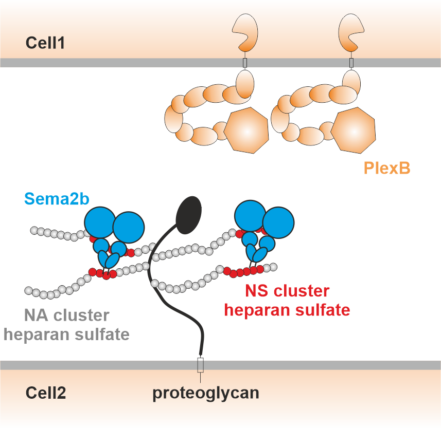
In addition to binding to plexins, semaphorins also interact with sugars on the cell surface. The research team investigated the molecular mechanisms of this interaction.
"We found that some semaphorins bind to sugars on the cell surface, especially negatively charged sugars. The higher the charge of the sugar, the stronger the binding. We also found that these sugars bind to a small part of the semaphorin, a little tail that is positively charged. Interestingly, when we removed this tail using protein engineering methods, the axons turned right instead of left in certain areas. This suggests that we could influence the growth and direction of axons using sugars," explains Farah Nourisanami, an Iranian PhD student at the Faculty of Science at Charles University.
The implications of these findings offer potential avenues for therapeutic interventions in neurodegenerative diseases and nerve injuries. "Some semaphorins inhibit the regeneration of damaged nerve tissue. Understanding their interaction with cell surface sugars could play an important role in the development of treatments for conditions caused by brain and spinal cord injury," said Daniel Rozbesky, Head of the Laboratory of Structural Neurobiology (Institute of Molecular Genetics of the Czech Academy of Science and the Faculty of Science of the Charles University) at the BIOCEV Research Centre in Vestec near Prague.

Proposed mechanism by which semaphorins (blue) bind to sugars (white spheres) on the cell surface and are thus presented to plexin receptors (orange) on surrounding axons. Source: PNAS
Contact:
Daniel Rozbeský
BIOCEV
daniel.rozbesky@natur.cuni.cz
Read also
- Scientists discover new species of rare fungi thanks to arsenic analysis
- MICAL1 plays a key role in cellular dynamics by controlling the cytoskeleton
- Groundbreaking study maps brain’s recovery process after stroke
- IOCB Prague expands overseas, opens a new branch in Boston
- Speedier and more effective treatment of serious illnesses
- Scientists from IOCB Prague help to improve medical drugs
- Newly discovered regenerative cells may revolutionize wound healing
- Genetic mixing as a path to survival
- Powerful winter lightning discharges triggered prolonged whistling around the Earth
- Czech discovery writes a new chapter in electrochemistry textbooks
Contacts for Media
Markéta Růžičková
Public Relations Manager
+420 777 970 812
Eliška Zvolánková
+420 739 535 007
Martina Spěváčková
+420 733 697 112