How to repair the spinal cord: Kristýna Kárová explores nerve cell recovery
16. 07. 2024
In the Czech Republic alone, up to 300 people suffer spinal cord injuries every year. The aftermath of damaging this delicate bundle of nerve fibers is generally irreversible. Or is it? Kristýna Kárová from the Institute of Experimental Medicine at the Czech Academy of Sciences is focusing intensely on the possibilities of axon regeneration following spinal cord injuries. An article about her research was published in the quarterly of the Czech Academy of Sciences, A / Magazine.
When you need to scratch your nose, your brain evaluates the situation and sends a signal through the motor pathways of the spinal cord to the muscles in your hand, which quickly complete the task. Simultaneously, though, there’s a message traveling up to your brain via the sensory pathways of the spinal cord from your toes, informing it that they are starting to feel cold. Our control center then immediately sends further commands through the spinal tracts to the body to eliminate this sensation of cold.
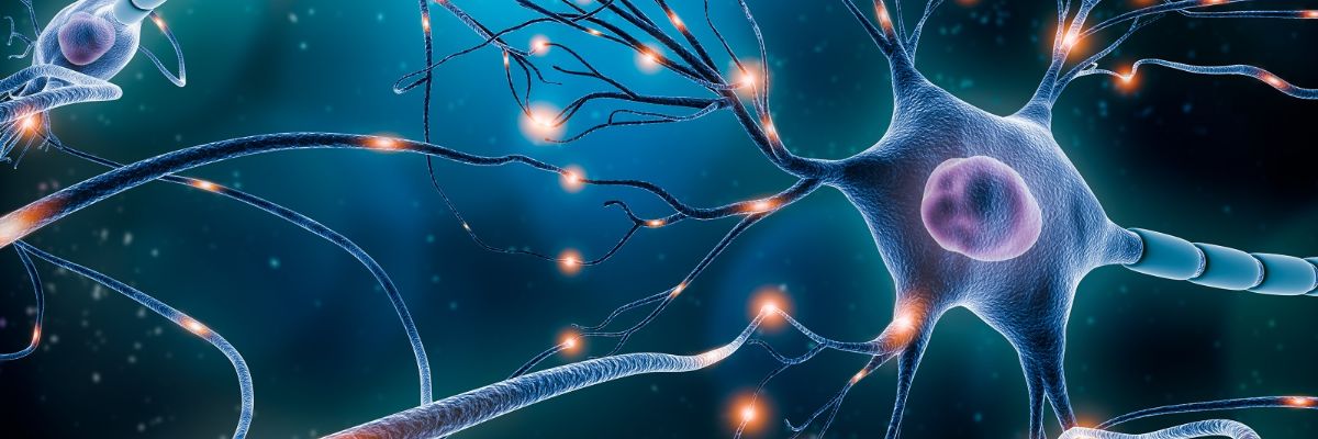
At any given time, there’s a lot happening inside the medulla spinalis. The nearly half-meter-long spinal cord, about as thick as a thumb, contains approximately thirteen-and-a-half million nerve cells – neurons – responsible for transmitting information. “We can picture a neuron as a hand. The palm represents the body of the cell, the fingers are dendrites, which are short extensions that receive signals. The forearm is like a long extension of the neuron called the axon, which connects to other neurons via synapses and transmits information,” explains neuroscientist Kristýna Kárová from the Department of Neuroregeneration at the Institute of Experimental Medicine of the Czech Academy of Sciences (CAS).

The spinal cord consists of 31 segments, each giving rise to a pair of spinal nerves that branch out to various parts of the body.
While a nerve cell can have thousands of dendrites, it always has only one axon. But it’s a big one – reaching up to one meter in length. Signals are transmitted between neurons thanks to a combination of electrical and chemical processes and travel through the spinal cord at speeds of up to 120 meters per second. But even on this sophisticated highway, a traffic jam can occur – when the spinal cord gets damaged, despite the protective efforts of the spine.
Wrong way: do not enter
Injury, various diseases, and tumors – the most common causes of spinal cord injury, also known as spinal lesions. At the site of the injury, the flow of information between the brain and the rest of the body is interrupted, often resulting in not only motor impairments but also sensory ones, or even disruptions to the autonomic nervous system, which controls the functions of internal organs. The higher the lesion is located, the more extensive the consequences.
“A clearly defined cavity forms in the spinal cord where the signal cannot pass through. A glial scar forms around the lesion, isolating the damage from the rest of the tissue and blocking the axons’ attempts to grow through the area,” Kárová describes.
As a result, the affected axons typically become inactive and their ends degenerate. However, the more dynamic axons sometimes try to save the situation and grow around the lesion. But their spontaneous efforts are not enough to restore the disrupted connections – at least not in humans.
That is why researchers everywhere have struggled for years to find ways to stimulate these inactive neuronal extensions to regenerate. Kristýna Kárová and her colleagues are also striving to initiate neuron renewal in the motor pathways of the spinal cord, aiming to restore the body’s original mobility. And they are doing this with the help of gene therapy.

Kristýna Kárová from the Institute of Experimental Medicine of the CAS. (CC)
The forgotten recipe
PI3K-delta. That is the name of a growth-promoting enzyme from the kinase group, which plays a crucial role in the development of the central nervous system. During the period when neurons are forming, this enzyme is abundant throughout the body. However, once nerve cells establish synapses and settle into the neural network, the production of this kinase naturally declines. Neurons simply transition from an active state to a more “settled” lifestyle, much like a teenager maturing into adulthood.
Back in 2007, German neuroscientists pondered this idea: “What if we reminded mature neurons of their youthful vigor and taught them how to produce the unbridled kinase again?” Interesting things started to occur in the cells stimulated by the enzyme. Researchers in Cambridge then embarked on kinase experiments with cell cultures, and years of experimentation proved that this protein promotes the regeneration of cortical neurons.
“We built on the findings of our international colleagues and tried to stimulate axon regeneration in the damaged spinal cords of rats with PI3K-delta. We essentially gave the neurons in the motor cortex of their brains the recipe for this ‘active’ protein, prompting the cells to start producing it again,” Kárová recounts.
The experimental results confirmed suspicions about the kinase’s power – Kárová’s team managed to activate a signaling pathway that supports regeneration in injured rodents and modulate the transport of molecules into the axons using PI3K-delta. In other words, the researchers forced the disabled axons to grow and reintegrate.

A cross-section of the spinal cord above the lesion (i.e., injury).
A virus in the role of messenger
The scientists delivered this crucial recipe to the rat test subjects with a simple injection. This allowed them to introduce a specially modified viral particle containing genetic information into the relevant part of the brain, convincing the neurons to produce the kinase.
“It’s a virus that doesn’t pose a threat to the animal because it cannot replicate itself. However, it can enter certain cells, so it basically works like an envelope we can use to send a message to the rat’s neurons,” the neuroscientist explains.
So, what exactly did the kinase activity cause in the rodents’ bodies? Histological analysis showed that under the protein’s influence, many new axons formed in the rats’ spinal cords. The damaged ones managed to overcome the injury by growing around it and lengthened by about one centimeter. Electrophysiological examinations also revealed that the growth of these extensions didn’t stop there – the axons successfully reintegrated into the neural network.
“When we stimulated the spinal tract using electrodes, we detected a signal below the lesion and even in the surrounding muscles. Simply put, the impulse overcame the obstacle of the injury and reached its destination,” Kárová clarifies.

Kristýna Kárová in her laboratory. (CC)
Sugar as motivation
Rats love sugar. Researchers exploit this fact to test whether and how the histologically documented “restart” of axons translates into improved motor abilities in these lab animals. Sweet rewards are prepared on steps at various heights or at the opposite end of a horizontal ladder with different-sized gaps between the rungs. The scientists then release the rodents into this sweet paradise and count how many sugar cubes they consume within a certain timespan or how skillfully they can navigate the ladder.
These tasks are first trained with healthy animals. Then, so-called behavioral tests are conducted on rodents with spinal cord injuries in the dorsal corticospinal tract that have received gene therapy.
“Injuries manifest in rats as deficits in their front paws. They aren’t completely paralyzed but have motor control issues. They tend to slip on the ladder or steps, fall through, lack balance, and struggle with fine grips... However, our tests show that within four months of administering the kinase recipe, their motor skills significantly improve,” Kárová reports.

The shape of the spinal cord gray matter resembles a butterfly.
Hope for the paralyzed?
After five years of concentrated efforts on the project, the neuroscientist’s team is now preparing to publish the promising results of their research. They are also evaluating a long-term study on the effects of the kinase in rats’ bodies a year after administering the therapy. This study also appears optimistic.
It’s no wonder that the successes of this research instill hope in people with spinal cord injuries. The International Foundation for Research in Paraplegia, based in Switzerland, has even financially supported it. However, Kárová remains cautious.
“Our approach does have potential, but we are developing an experimental treatment, so the journey to applying our findings to humans is still a very long one. It involves years of further studies, especially chronic studies on rats, followed by other animal species like monkeys or pigs. And even then, success is not guaranteed,” the researcher warns.

Another cross-section of the spinal cord. (CC)
Kárová is alluding to the fact that scientists everywhere have often found that what works well in animal models is not guaranteed to have the same effect in the human body.
“But without faith, this work wouldn’t be possible. So I sincerely hope that one day, we will manage to get a person with a spinal cord injury moving again,” the scientist concludes.
__________________________________
Mgr. Kristýna Kárová, Ph.D.
Institute of Experimental Medicine, Czech Academy of Sciences
Kristýna Kárová studied immunology at the Faculty of Science, Charles University (Prague), and completed her doctoral studies at the Second Faculty of Medicine at the same university. Since 2012, she has been working at the Institute of Experimental Medicine of the CAS. Her research primarily focuses on the potential use of gene therapy to stimulate axon regeneration following spinal cord injury. She has worked with many prestigious international institutions, such as New York Medical College, the University of Cambridge, and King’s College London. In 2023, she was awarded the Otto Wichterle Award for outstanding young scientists.
__________________________________
You can read the article in Czech in the quarterly of the CAS, A / Magazine, published by the Czech Academy of Sciences:
Written and prepared by: Radka Římanová, External Relations Division, CAO of the CAS
Translated by: Tereza Novická, External Relations Division, CAO of the CAS
Photo: Shutterstock; Jana Plavec, External Relations Division, CAO of the CAS; Kristýna Kárová’s archive
 The text and photos labeled CC are released for use under the Creative Commons license.
Read also
- Small but Mighty – The Enceladus Moon Is Flexing with Saturn’s Magnetic Field
- Light vs. Antibiotics – Can We Get Harmful Substances Out of Our Water?
- Better ECGs and Industrial Superlasers – Real-World Results of CAS Research
- Czech Academy of Sciences to launch a joint-stock company
- In the Age of AI, Spotting a Fake Photo Is Harder Than Ever, Expert Says
- How to Turn Ideas into Successful Grants: The New ERC Incubator Is Offering Help
- SciComm 360° Tackled How to Communicate Science in the Age of Disinformation
- The Academy of the Future? A New Vision for Attracting Scientific Talent
- When Cars Fly and Bullets Swerve – Physics Gone Wrong on the Silver Screen
- The Academy to Boost Excellence and Careers in Research with New Programs
The Czech Academy of Sciences (the CAS)
The mission of the CAS
The primary mission of the CAS is to conduct research in a broad spectrum of natural, technical and social sciences as well as humanities. This research aims to advance progress of scientific knowledge at the international level, considering, however, the specific needs of the Czech society and the national culture.
President of the CAS
Prof. Radomír Pánek started his first term of office in March 2025. He is a prominent Czech scientist specializing in plasma physics and nuclear fusion.